Lab Experience at the Juntendo University Hospital – Linda Guo
Personal reflections 2nd lab rotation
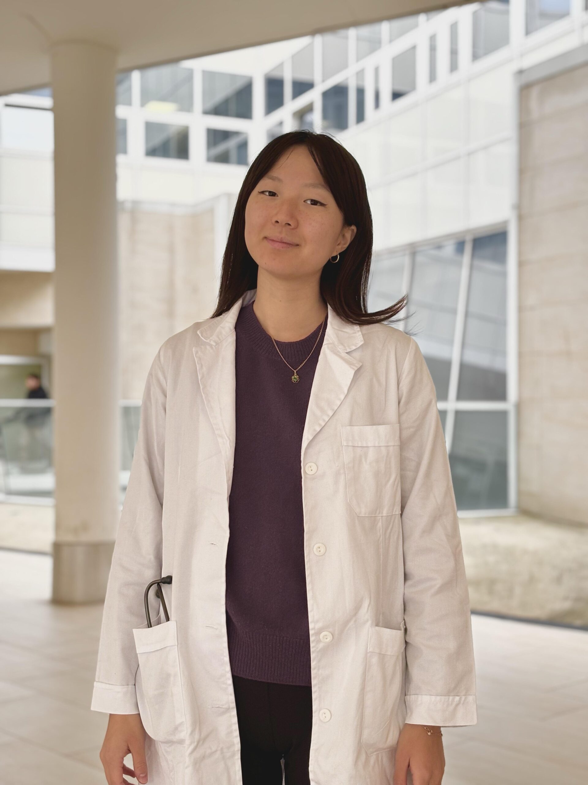
Linda Guo, Virgilio 6° Cohort Student, UNIMI
Name: Linda Guo
Mentor: Prof. Filippo Martinelli Host PI: Prof. Takenori Inomata
My second lab rotation at Juntendo University Hospital and the Inomata Challenging Lab has been a transformative academic and personal journey. Over the course of six weeks, I was exposed to a rich combination of clinical practice, experimental laboratory techniques, and systematic review methodology. This immersive experience not only deepened my understanding of ophthalmology but also strengthened my confidence in pursuing translational research with clinical relevance.
From the outset, I was impressed by the advanced and structured clinical environment in Japan. I participated in various outpatient clinics focused on general ophthalmology, dry eye disease (DED), and contact lenses.
Observing the evaluation and management of ocular surface disorders in Japan provided a valuable comparison to Italian clinical practice. For instance, I learned that treatments like Diquafosol and Mucosta—commonly prescribed for DED in Japan—are not available in Italy. I also witnessed the routine use of nasolacrimal duct plugs, which is rare in my home hospital. These observations broadened my perspective on international therapeutic approaches.
One of the most exciting aspects of this rotation was the access to rare surgical procedures and training opportunities. I had the unique chance to observe a corneal transplant, a procedure that I had not yet seen during my clinical years in Italy. Witnessing the precision and delicacy required for penetrating keratoplasty gave me a renewed respect for corneal surgery and its impact on vision restoration.
Even more groundbreaking was the opportunity to attend Japan’s first robotic retina surgery using the CNA0T0 system—a minimally invasive, robot-assisted procedure that integrates endoscopy to visualize the anterior retina. This was an incredibly rare experience, not currently available in my home university, and it gave me insight into the future of microsurgical technology and its potential applications in complex vitreoretinal procedures.
In addition to observation, I was able to practice cataract surgery hands-on using pig eye models on two occasions. Practicing the steps of phacoemulsification—such as capsulorhexis, hydrodissection, and intraocular lens implantation—was invaluable for building confidence and familiarity with surgical instrumentation. These sessions were an important bridge between theory and practice, and something I had not previously had the chance to do in my home country.
The lab component of my rotation focused primarily on flow cytometry and RNA extraction from mouse ocular tissues. Initially, I encountered a setback during RNA extraction from cornea, conjunctiva, and lymph nodes: despite following standard protocols, the RNA yields were consistently below 200 ng, insufficient for reliable downstream applications like qPCR.
Instead of being discouraged, this challenge became one of the most instructive parts of my rotation. After discussing the issue with the lab team, we hypothesized that room temperature handling could be leading to RNA degradation. To test this, we designed a comparative experiment using tissues kept on ice versus room temperature samples. The result was clear—iced tissues produced significantly higher RNA yields and better purity, confirming the importance of temperature control during tissue collection and processing.
This hands-on troubleshooting reinforced my understanding of experimental design and taught me the value of hypothesis-driven adjustments. It also showed me that even routine protocols require careful adaptation to specific tissues, particularly when working with small and delicate samples like the cornea.
Parallel to the clinical and lab activities, I also worked on a systematic review project under the guidance of Ken-san, an expert in meta-analysis. The review focused on optimal storage conditions for Platelet-Rich Plasma (PRP) and the preservation of growth factor activity. I screened over 2,000 abstracts, extracted data from over 130 full texts, and helped organize the results based on storage temperature, duration, and type of growth factor assays.
Through this project, I learned how to apply PRISMA guidelines, evaluate study quality, and construct evidence tables. It enhanced my skills in critical reading, scientific writing, and data synthesis—skills that will be vital as I work toward future publications. This was my first real experience working on a formal academic review from conception to drafting, and I am grateful for the mentorship and structure provided by the lab
Equally important to the technical training was the mentorship and lab environment itself. Prof. Inomata and the lab members created a welcoming and intellectually stimulating atmosphere. I felt respected and encouraged to ask questions, participate in discussions, and share my observations. Regular meetings and informal conversations helped me integrate more deeply into the research group, despite the language barrier at times.
Attending the Fujiretina Conference added another dimension to my rotation. It allowed me to hear about cutting-edge innovations in imaging and surgical techniques, and to meet ophthalmologists and researchers from Japan, the U.S., and Europe. The focus on collaboration and real-world clinical challenges resonated strongly with my own aspirations as a future clinician-scientist.
Conclusion
Looking back, this rotation provided me with invaluable insights that I would not have had access to in my home institution. Observing robotic surgery, practicing cataract surgery on pig eyes, and working hands-on with RNA and flow cytometry protocols have all contributed to shaping a clearer vision for my future career. I now feel better equipped to bridge the gap between clinical medicine and scientific research.
Furthermore, this experience solidified my interest in ocular surface disease, corneal immunology, and experimental therapeutics. The knowledge and confidence I gained here will support my continued development, both in clinical settings and in the lab.
This rotation at Juntendo University Hospital has been one of the most formative experiences of my training. It challenged me, inspired me, and equipped me with skills that go far beyond the textbook. I return with not only new technical abilities, but also a deeper appreciation for global collaboration, translational research, and surgical innovation. I am sincerely thankful to Prof. Nakao, Prof. Inomata, Prof. Martinelli, and all the lab members who made this opportunity so impactful.